Prof. Ralph Müller
Exploring the Mechanoregulation of Bone Regeneration
In over 100 years, the remarkable ability of bone to adapt to its mechanical environment has been a source of scientific fascination. Bone regeneration has been shown to be highly dependent on the mechanical environment at the fracture site. It has been demonstrated that mechanical stimuli can either accelerate or impede regeneration. Despite the fundamental importance of the mechanical environment in influencing bone regeneration, the molecular mechanisms underlying this phenomenon are complex and poorly understood.
Keywords
Bone, Mechanobiology, Spatial transcriptomics, Gene expression, Finite element modelling, Image processing
Labels
Semester Project , Internship , Bachelor Thesis , Master Thesis
Description
Goal
Contact Details
More information
Published since: 2026-06-19 , Earliest start: 2026-01-19 , Latest end: 2026-08-31
Organization Müller Group / Laboratory for Bone Biomechanics
Hosts Mathavan Neashan
Topics Medical and Health Sciences , Engineering and Technology
Development of a Heterocellular Human Bone Organoid for Precision Medicine and Treatment
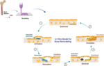
Our goal is to establish a heterocellular 3D printed bone organoid model comprising all major bone cell types (osteoblasts, osteocytes, osteoclasts) to recapitulate bone remodeling units in an in vitro system. The organoids will be produced with the human cells, as they could represent human pathophysiology better than animal models, and eventually could replace them. These in vitro models could be used in the advancement of next-generation personalised treatment strategies. Our tools are different kinds of 3D bioprinting platforms, bio-ink formulations, hydrogels, mol-bioassays, and time-lapsed image processing of micro-CT scans.
Keywords
3D printing, bone organoids, co-culture, bioreactor, hydrogels, drug testing
Labels
Semester Project , Internship , Bachelor Thesis , Master Thesis , ETH Zurich (ETHZ)
Description
Goal
Contact Details
More information
Published since: 2026-06-01 , Earliest start: 2026-07-01 , Latest end: 2027-07-01
Organization Müller Group / Laboratory for Bone Biomechanics
Hosts Steffi Chris
Topics Engineering and Technology , Biology
Spatial Proteomics of Mechanically-Driven Bone Healing

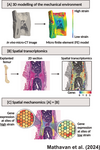
Bone healing is profoundly influenced by its mechanical environment. Advances in spatial proteomics now allow us to map protein expression within intact tissue and directly relate it to local biomechanical cues. The Laboratory for Bone Biomechanics is developing a new line of research within spatial mechanomics (DOI: 10.1126/sciadv.adp8496), integrating spatially resolved proteomic data with in silico models of the mechanical environment at fracture sites. This approach enables us to investigate, at cellular resolution, how mechanical forces shape protein-level signalling during bone repair.
Keywords
Bone, Mechanobiology, Spatial Proteomics, Protein Expression, Aging, Sex Differences, Mechanical Loading, Finite Element Modelling, Image Analysis
Labels
Semester Project , Internship , Bachelor Thesis , Master Thesis
Description
Goal
Contact Details
More information
Published since: 2026-04-28 , Earliest start: 2025-12-01 , Latest end: 2026-12-31
Organization Müller Group / Laboratory for Bone Biomechanics
Hosts Mathavan Neashan
Topics Medical and Health Sciences , Engineering and Technology , Biology